Resting-state fMRI
Resting-state fMRI (rsfMRI) is a method of functional brain imaging that can be used to evaluate regional interactions that occur when a subject is not performing an explicit task. This resting brain activity is observed through changes in blood flow in the brain which creates what is referred to as a Blood Oxygen Level Dependent (BOLD) signal that can be measured using functional Magnetic Resonance Imaging (fMRI).
Because brain activity is present even in the absence of an externally prompted task, any brain region will have spontaneous fluctuations in BOLD signal. Resting-state functional connectivity research has revealed a number of networks which are consistently found in healthy subjects and represent specific patterns of synchronous activity. However, the mechanisms governing the temporally coherent spontaneous fluctuations in BOLD signal remain unclear. Exploration of these mechanisms is one of the research interests in BISP Lab.
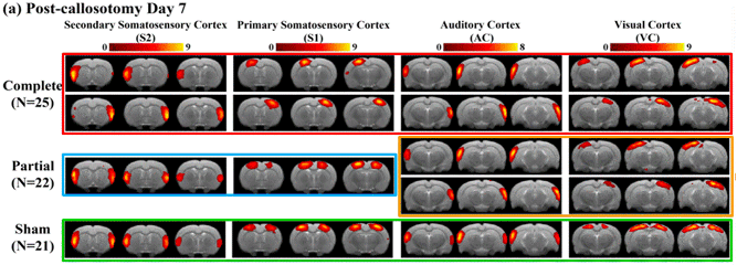
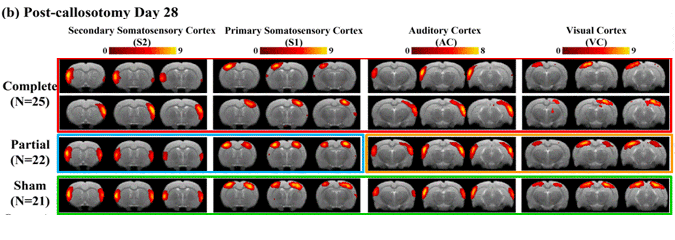
One of our projects, entitled “Brain resting-state functional MRI connectivity: Morphological foundation and plasticity”, directly demonstrates that axonal connections are the indispensable foundation for rsfMRI connectivity and that such functional connectivity can be plastic and dynamically reorganized atop the morphological connections.
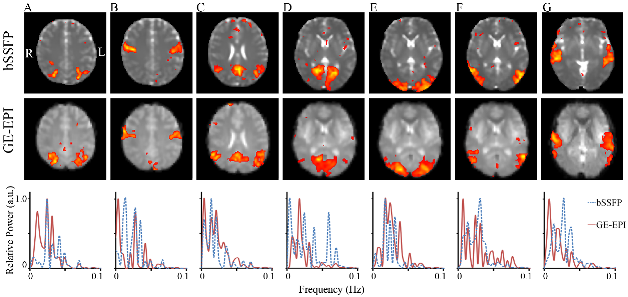
Another representative study investigated feasibility of passband balanced steady-state free precession (bSSFP) imaging for distortion-free and high-resolution rsfMRI.