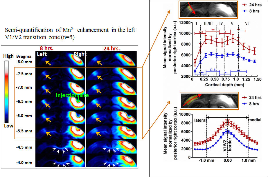
Typical T1- and T2-weighted images, T1 and T2 maps from one animal at 0 and 4 weeks after CCl4 insult. Typical measurement ROIs are shown in T1 and T2 maps.
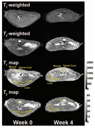
Microbubbles
(a) Representative light microscopy of MION-entrapped PMBs. (b) Histogram showing diameter distribution for a representative batch of MION-entrapped PMBs. The mean diameter of MION-entrapped PMBs is 9.77 μm.
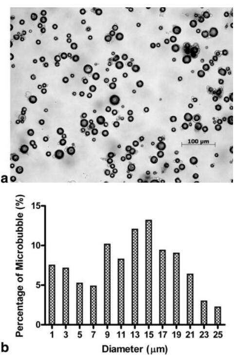
The typical multiecho GE signals from a MION-free PMB phantom (5% volume fraction) in (a) its initial well-suspended state and (b) its final microbubble-free state with the monoexponential fitting in dotted lines. (c) R2* vs time. The multiecho GE signals from a MION-entrapped PMB phantom (5% volume fraction) in (d) its initial well-suspended state and (e) its final microbubble-free state. f: R2* vs time.