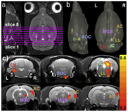
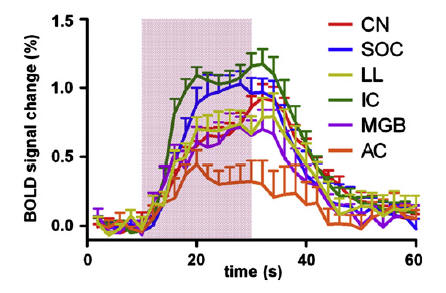
Activation (r>0.2) using broadband noise stimulus can be observed in multiple auditory structures: cochlear nucleus (CN), superior olivary complex (SOC), lateral lemniscus (LL), inferior colliculus (IC), medial geniculate body (MGB) and auditory cortex (AC).
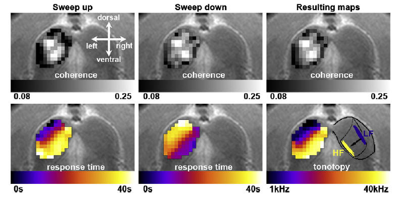
High fidelity tonotopic mapping using swept source fMRI.
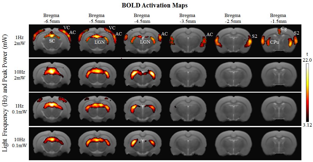
First visual fMRI study that demonstrated large-scale brain activation using strong low frequency light stimulation, such stimulus evoked responses in the visual system and brain regions that play a role in multisensory interaction and attentional modulation of sensory processing. Our current study provides a functional understanding to cortical cross-modal activity and its influences on the subcortex upon visual stimuli of different intensities and frequencies.
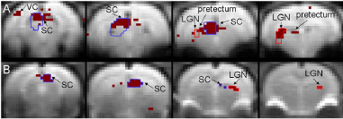
Gradient-echo and spin-echo activation maps following short duration visual stimulation. (A) Activation map computed by applying Analysis of Variance on the four slice fMRI data (slices 1 to 4 arranged from left to right) of a representative animal scanned with the gradient-echo (GE) sequence. Voxels with p<10-3 are colored dark red. The blue and red regions of interest (ROIs) cover voxels containing the contralateral superior colliculus (SC) and lateral geniculate nucleus (LGN), respectively. The contralateral hemisphere is on the left. (B) Activation map computed from the average fMRI data of all animals scanned with the spin-echo (SE) sequence. Voxels with p = 0 (beyond computer precision) are colored dark red. Blue and red ROIs cover such voxels in the contralateral SC and LGN, respectively. The contralateral hemisphere is on the right. Low p-value regions of the SC, LGN, pretectum, and visual cortex (VC) are labeled.