Biomedical imaging has come to play a key role in clinical diagnostics, as well as basic life science research. In particular, early detection of rare cancer cells and understanding their behaviours with high efficiency and accuracy has been a daunting task in the arena of biomedicine. The key technological challenge is to pinpoint individual cells in a large population and to analyse them one by one.
Now a multidisciplinary team from HKU, led by the Faculty of Engineering, has come up with new technology that will not only drastically speed up the rate at which individual cells can be monitored, but also makes cellular resolution much clearer thereby making detection of suspect cells easier.
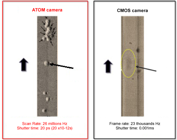
Called ATOM – Asymmetric-detection Time-stretch Optical Microscopy – it can capture images of moving cells up to 10,000 times faster than existing camera technologies.
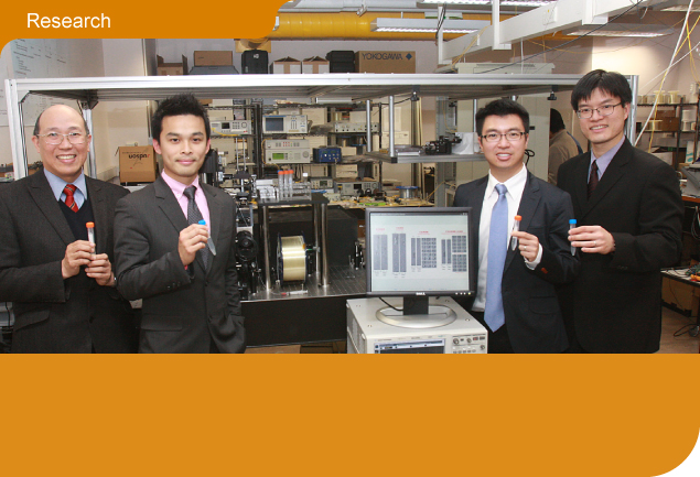
Head of the crossdisciplinary team that developed ATOM is Dr Kevin Tsia Kin-man, who is affiliated with the Department of Electrical and Electronic Engineering and also teaches in the Medical Engineering programme.
I’m a strong advocate for turning research output into something practical and useful. This is particularly relevant to biomedical engineering as it is the main driving force for better health care.
Dr Kevin Tsia Kin-manInnovative camera concept
“There are two key features to the new technology,” he said. “One, we’re not using current camera technologies, as they are what limits the imaging speed. We have developed a system that functions as a camera, but is not the usual concept of a camera.
“And two, a pulse laser is the source for imaging – the pulsing speed of the flashlight is very fast so that is the frame rate, 1,000 times faster than a conventional camera. Using an optical encoding technique, we are able to store the image data into the pulse of the flashlight – one pulse carries one image frame.”
The implications of ATOM for improving flow cytometry (the gold standard for cell analysis), both in terms of speed and accuracy of detection, are great. “Flow cytometers for instance can test blood cells, but there is a lot of room for human error in the blood cell screening and the subsequent data analysis processes,” said Dr Tsia. “The main reason is the lack of image information for each cell in standard flow cytometers. And it is generally true that accessing the image data of each cell facilitates better cellular identification/discrimination and thus yields high-confidence statistical data.”
The need for enabling imaging capability in flow cytometers has already resulted in the recent development of Imaging Flow Cytometers. However, they use a conventional camera so the speed is limited to about 1,000 cells per second.
“We’re marrying the super fast camera system with flow cytometer technologies,” said Dr Tsia. “The result is biomedical imaging technology that provides not only the best combination of high-contrast and high-speed single cell imaging, but also – because it can generate an enormous image data within a short period of time – a new paradigm in high-throughput cell screening, big data bioimaging.”
ATOM can capture images of ultra-fast-moving living cells with cellular resolution in flow at a speed as high as 10 metres per second which translates to an imaging throughput of 100,000 cells per second, more than 1,000 times faster than any existing charge-coupled device (CCD) or complementary metal oxide semiconductor (CMOS) camera technologies.
Dr Tsia came to HKU in 2009 as an Assistant Professor. “I was doing my PhD at the University of California, Los Angeles (UCLA). My interest is optics in general, and originally my research was in telecommunications – fibre optics for the internet. But I switched midway because I realised the optical technologies in telecommunication can be borrowed and adapted to advance optical bioimaging. This is a relatively underexploited area, and thus motivated me to move into biomedical engineering.”
The team which developed ATOM has come together gradually over the past five years. Key members are Dr Anderson Shum Ho-cheung from Mechanical Engineering, Dr Kenneth Wong Kin-yip from Electrical and Electronic Engineering and Professor Godfrey Chan Chi-fung, Tsao Yen-Chow Professor in Pediatrics and Adolescent Medicine, from the Faculty of Medicine.
Multiple applications
The HKU team is focussing on the biomedical applications, but the technology has other applications. It could be used in industries such as paper manufacturing for surface inspection, and for high-speed quality checks in computer chip manufacture.
“We’re now working on validating and optimising the systems – the next stage is working with doctors on the diagnostics side,” said Dr Tsia. “There is an immediate need for this in the market, so we are patenting the technology.
“I’m a strong advocate for turning research output into something practical and useful. This is particularly relevant to biomedical engineering as it is the main driving force for better health care. I tell my students research is not just about sitting in a laboratory – our ideas should be used out there in the real world to improve medical care.” ■
 Compared to conventional complementary metal oxide semiconductor (CMOS) cameras, ATOM can capture images of ultra-fast-moving living cells with cellular resolution in flow at a speed as high as 10 metres per second.


